General
Life
Accessibility Options

General

General Products
Simple & Transparent! Policies that match all your insurance needs.


Scan to download

Life

Life Products
Digit Life is here! To help you save & secure your loved ones' future in the most simplified way.

Scan to download

Claims
Claims
We'll be there! Whenever and however you'll need us.

Scan to download
 General
 Life
Life
Renewals
Resources
Resources
All the more reasons to feel the Digit simplicity in your life!

Scan to download
Our WhatsApp number cannot be used for calls. This is a chat only number.


Sonography, commonly known as ultrasound, is more than checking foetal growth during pregnancy. It helps doctors identify underlying issues with your vessels, tissues, and organs without requiring an incision.
This article will explain all about what sonography is, the types, precautions, and risks associated with the same. Read on to know more!
An ultrasound or sonography is a safe and non-invasive clinical examination method that uses high-frequency sound waves to record images inside your body. For instance, sonography effectively monitors the progress in pregnancy in obstetrics and assists in identifying heart function and physiology in cardiology.
It also visualises tumours and how they respond during treatments in oncology. In addition, unlike other imaging methods, sonography includes no ionising radiation.
Besides knowing what sonography is, you must understand that there are various techniques to diagnose different body conditions. Here are the different types of sonographies and their uses for each:
Much like a gynecologic exam, the sonohysterography or hysterosonography method uses sound waves to capture images inside a woman’s uterus.
Uses of Hysterosonography: It helps to diagnose miscarriages, infertility, and unexplained vaginal bleeding.
This sonography test uses sound waves to capture images of an embryo or a foetus in a pregnant woman.
Uses of Obstetric Ultrasound: It is considered a preferred method to monitor pregnancy and the child's development inside a mother’s womb.
This method captures images of the structures in your upper abdomen.
Uses of Abdomen Ultrasound: It assists in diagnosing abdominal pain or enlargement and examines the functioning of kidneys, gallbladder, liver, pancreas, spleen, bile ducts, and aorta.
In this method, the sound waves capture pictures of the internal structures of a breast.
Uses of Breast Ultrasound: Doctors conduct this method to diagnose breast lumps and other abnormal structures found while performing a breast MRI or a mammogram.
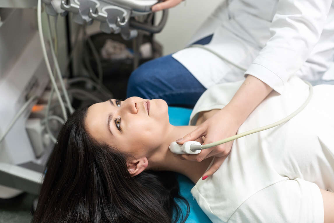
Carotid sonography uses sound waves to capture images of the carotid arteries in your neck. This artery carries blood from your heart to your brain. Besides, doppler technology is also part of this exam.
Uses of Carotid Ultrasound: This method is used to diagnose narrowing or blockage of carotid arteries (stenosis) that increases the chances of stroke.
This method captures images of your body's muscles, ligaments, nerves, tendons, and joints.
Uses of Musculoskeletal Ultrasound: It helps to diagnose strains, arthritis, sprains, tears, trapped nerves, and related musculoskeletal conditions in a body.
Pelvic sonography captures pictures of the organ structure in your pelvis and lower abdomen. Moreover, there are three different types of pelvic sonography which include- vaginal (for women), abdominal, and rectal (for men).
Uses of Pelvic Ultrasound: Doctors use this method to diagnose issues in your body's urinary and reproductive systems.
This method produces images of the prostate gland.
Uses of Prostate Ultrasound: It assists in diagnosing difficulty while urinating. Additionally, this method helps to detect a nodule found during a rectal exam, an enlarged gland, abnormal structures, or an elevated blood test result.
This method uses sound waves to capture images of the testicles and the surrounding tissues of a male.
Uses of Scrotum Ultrasound: It is a primary procedure that allows the evaluation of testicle disorder, epididymis, or the tubes precisely next to the testicles and scrotum.
This method uses sound waves to capture images of the thyroid gland in your neck.
Uses of Thyroid Ultrasound: Doctors conduct this method to examine nodules or lumps while evaluating a routine physical exam or related imaging test.
This method uses sound waves to capture pictures of the veins in your body.
Uses of Venous Ultrasound: It commonly helps to diagnose blood clots, especially in the veins of your leg. Also, it examines deep vein thrombosis effectively.
It is essential to learn the way it works to know about sonography tests thoroughly:
Step 1: In sonography, an instrument known as a transducer releases high-frequency inaudible sound waves.
Step 2: These sound waves travel steadily inside your body until they hit a dense object such as a bone or an organ.
Step 3: Then, depending on the tissue present, these waves either continue to travel further or get reflected.
Step 4: The sound waves that reflect or echo get communicated to the ultrasound device.
Step 5: Depending on the return time of each echo and the speed of sound waves in tissues, the imaging device calculates the distance. It is between every structure and the probe.
Step 6: The intensity and distance of all echoes are modified into a 2-dimensional image. This appears on the sonography imaging screen.
Sonographers and ultrasound specialists are well-versed in performing this test, after which a doctor or a radiologist interprets the images.
Every medical test and examination come with its own set of rules that both you and your doctor follow. However, depending on the doctor's suggestion, below are the things you must follow to prepare for the test:
The procedure for sonography is simple and continues for only a few minutes. Here is the process of the test:
Step 1: Before the sonography exam, your doctor will ask you to remove any jewellery you wear to examine.
Step 2: Next, you will have to change into a gown so they can examine the area easily.
Step 3: The staff will ask you to lie on an examination table.
Step 4: Lastly, to start the process, your sonographer might cut or remove some of the clothing from the examination area.
Step 5: A sonographer will apply a lubricating gel over the examination area to prevent air pockets from blocking sound waves that create images. This gel is easy to remove and doesn’t cause side effects.
Step 6: A sonographer will press a transducer against the examination area and move it as required to capture pictures of that area.
Step 7: The device sends sound waves in your body and gathers the ones that reflect. This, when sent to the computer, creates pictures.
Step 8: In some instances, doctors conduct an ultrasound inside your body by attaching a transducer to the probe and inserting it into a natural opening in your body.
Step 9: After the procedure, the sonographer cleans off the gel on your skin, and you can resume your normal activities.
Step 10: After conducting sonography, your medical expert will review all the images captured during the process and detect any abnormalities in your body.
Step 11: They might discuss reports and findings with you. Furthermore, you might need to undergo a CT scan, an MRI, or a biopsy tissue sample if there are any abnormalities.
Step 12: After your doctor diagnoses your condition based on these reports, they will start your treatment accordingly.
During the process, you might be required to change positions as per the technician’s instructions so that they can have better access to the examination area. This whole procedure lasts for 30 minutes or less than that, depending entirely on the area of examination and the type of ultrasound done.
It is important to note that during a Transesophageal Echocardiogram, the professional may insert a transducer inside your oesophagus. It is to collect images of your heart. Moreover, this process usually takes place under sedation. During the Transrectal method, a technician places a transducer inside the rectum. Additionally, in the transvaginal procedure, it goes inside a vagina.
Sonography, generally, doesn't include any risk, adverse effects, or complications. Moreover, since it is devoid of radiation, you also have no fear of side effects.
However, there might be few chances of raising the temperature of surrounding tissues in the examination area. Also, you must only undergo this test under the guidance of a trained practitioner.
Ultrasonography ensures a better and more detailed view of the functioning of organs, muscles, bones, etc. The benefits of sonography include the following:
Following are the precautions both you and a sonographer require to maintain:
So, now you know what is sonography and its benefits, and the easy process it includes. First, however, ensure only a skilled professional is conducting this process, delivering prescribed doses, maintaining all safety measures and adhering to guidelines.
Protect What Matters - Explore Other Insurance Options