What is MRI Scan: Meaning, Working, Procedure & Advantages

MRI, also known as magnetic resonance imaging, is a standard scan in the medical arena all over the world. MRI scan uses a strong magnetic field and radio waves to create images of tissues and organs of the body. Also, since its invention, medical professionals and scientists have been in a continuous effort to make it more efficient and effective.
What Is an MRI Scan?
MRI, or magnetic resonance imaging, is a non-invasive imaging technology which is utilised to produce high-quality 3-D anatomical images. MRI scanners use radiofrequency energy (radio waves) and magnetic fields in these images. It is generally used for disease detection, treatment monitoring and diagnosis.
Precisely speaking, it is a nuclear magnetic resonance (NMR) that can also be used for other NMR applications (For example, NMR spectroscopy) besides medical applications.
How Does an MRI Scan Work?
An MRI primarily consists of two powerful magnets, which are this machine's three essential parts.
- Initial Phase: In general, the human body is primarily composed of H2O molecules, which comprise hydrogen and oxygen atoms. At the centre of each molecule is a proton that serves as a magnet and can be susceptible to any magnetic field. The molecules are usually randomly arranged in the human body, and they align in one direction after entering the MRI scanner.
- Middle Phase: Then, the second magnetic field is switched on, sending quick pulses that change every hydrogen atom's direction. After that, it immediately turns back to its original random state when switched off.
- Last Phase: After that, electricity is passed through gradient coils that make the coils vibrate, generating a magnetic field, which creates a knocking sound. However, the patient cannot detect these changes in their body, but the scanner detects those. Lastly, with the help of computers, it creates a detailed image that helps doctors to diagnose health issues.
How is the Procedure of MRI Scan Performed?
The procedure of MRI Scan is done in the following ways:
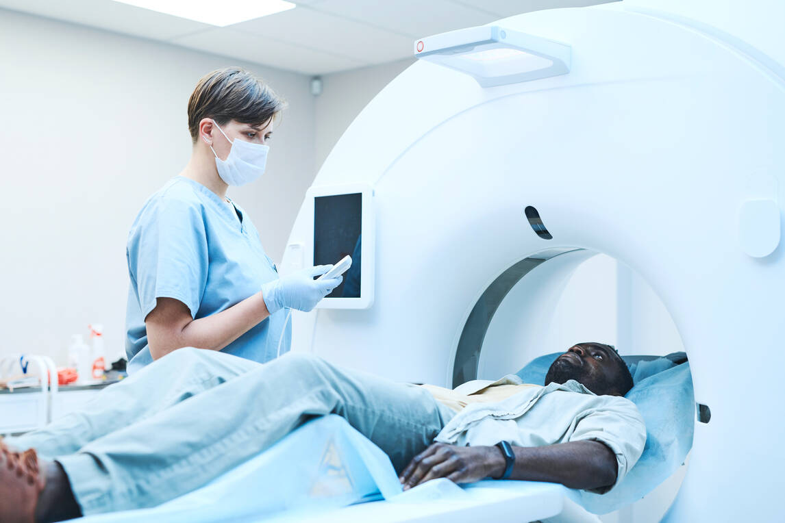
Phase 1 - During an MRI, you will lie straight on your back, which will be moved to the scanner. Also, you will be advised to lie, depending on your concerned body part. In most medical facilities, an MRI scanner is operated by a radiographer trained to perform imaging investigations.
Phase 2 - These radiographers control the machine with a computer in a different room to keep it safe from magnetic radiation. Also, you will be able to hear and talk with the medical professional monitoring you through the viewing window in the scanner.
Phase 3 - During the procedure, the machine might make a tapping sound resulting from the electric current going on and off. They can provide you with headphones and earplugs to wear to keep you still during the scan. In a few circumstances, you might also need to hold your breath for a few seconds. Regardless, depending on the size of the area to be scanned, the total scan can take 15 to 90 minutes.
What Happens During and After the Procedure?
After switching on the scanner, the concerned person will communicate with the patient via telecom to make them more comfortable. Also, they will start the machine once the patient is ready.
During the Scan
While the scan is running, it is essential to be still. Any movement during the scan can disrupt the image, equivalent to the shaking of a camera while taking a photo. It is essential to note that loud noises may come from the machine, which is normal. However, you might need to hold your breath depending on the scan.
In a few instances, the doctor will inject you with a contrast material (for example, gadolinium) in your arm through an intravenous line. This material magnifies a few details and doesn't usually cause an allergic reaction. But, of course, if you feel discomfort, you can always request the medical personnel to stop the process.
After the Scan
Upon completion of the scan, a radiologist will view the images, and if they are satisfied with them, they will be released. After, they will prepare a detailed report with a doctor, and you will be given an appointment to discuss your issue further.
Who Interprets the MRI Results and How do I get them?
Radiologists will typically analyse your MRI images and report the details to your doctor. After that, you will be provided with the report and other details. But, the time might depend on several factors, even if you are in an emergency.
What Are the Advantages of MRI?
A few of the advantages of MRI include the following:
- Zero Radiation Risks: MRIs have no such risks compared to X-Rays, where you are exposed to radiation. Some of the consequences of this exposure include vomiting, nausea, infection risk, etc. So, while you are choosing an MRI, you are not exposed to any of those risks.
- Clear Pictures: MRI scan produces clear pictures compared with other medical imaging gadgets. Moreover, it can even produce a clearer image of soft tissues within the body, allowing you to diagnose your disease better.
- Not Invasive: One of the issues with a few imaging techniques is their invasiveness. But, with MRI, you can be stress-free as the process is non-invasive.
- Captures Wide Pictures: One of the issues with X-Ray is that they can only take pictures of a particular body part. But, in the case of MRI, it can produce pictures of the whole body and a particular body part.
What Are the Disadvantages of MRI?
The side effects of an MRI are implausible. However, in a few instances, the contrast dye may trigger headaches, nausea, and burning or painful sensation at the point of injection. Additionally, allergic reaction to it is rare but can cause itchy eyes or hives.
What Is MRI Scan Used For?
MRI scans are used to examine your tissues, organs and skeletal system. However, depending on the requirement for medical needs, doctors will advise or prescribe for MRI.
Hence, it is evident that MRI scans are unequivocally helpful for medical sciences. Also, an MRI scan aids in diagnosing complex medical problems while resulting in faster treatments.
Protect What Matters - Explore Other Insurance Options


